Pelvic varices embolisation
Minimally invasive procedure performed by interventional radiologists to treat varices in the pelvic region. Carried out under precise radiological guidance by an expert team. Dr Nicolas PANGON, Consultant in interventional radiology, Centre Aquitain d'Imagerie & Bordeaux University Hospital, 06/02/2025.
Looking for answers based on your symptoms?
Read our patient guide: Chronic pelvic pain (women)What is it?
Pelvic varices embolisation is a minimally invasive procedure performed by interventional radiologists to treat varices in the pelvic region, which may cause chronic pelvic pain and other bothersome symptoms. This technique involves occluding the dilated veins responsible for blood flow into the varices.
Pelvic varices correspond to venous dilatation, more or less associated with reflux, which may affect the ovarian, peri-uterine, peri-vaginal and perineal veins (including vulvar veins). They may extend into the venous circulation of the lower limbs.
They may be responsible for various symptoms:
- Pelvic congestion syndrome, characterised by chronic pelvic pain of variable intensity, related to menstruation, which may be positional and worsened at the end of the day. These symptoms can be very disabling in daily life and may affect approximately 1 in 20 women.
- Lower limb varices, manifesting as leg pain, fed by pelvic reflux.
What causes them?
Pelvic varices occur when veins in the pelvic region dilate and enlarge. Several factors may lead to this situation:
- Pregnancy: Each pregnancy increases the risk of pelvic varices due to increased blood volume and pressure on the veins. Symptoms most often appear after the second pregnancy.
- Family history: Venous insufficiency may have a genetic component.
- Anatomical abnormalities of pelvic veins: rare compression syndromes may explain the symptoms.
How is the diagnosis made?
Diagnosis is both clinical, based on the medical history and physical examination, and supported by complementary investigations:
- Doppler ultrasound: may reveal dilatation of peri-uterine veins
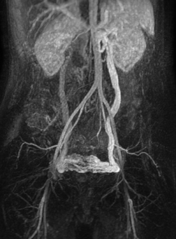
- Pelvic MRI: the reference examination, which also helps rule out differential diagnoses (endometriosis, fibroids, adenomyosis, etc.). It confirms dynamic reflux, particularly of the ovarian veins.
How does embolisation proceed?
Pelvic varices embolisation may be performed under local anaesthesia or sedation, as a day-case procedure. After a post-procedure observation period, patients may return home the same day. The procedure generally proceeds as follows:

- Vascular access: A catheter is inserted into a vein, usually in the groin at the level of the femoral vein.
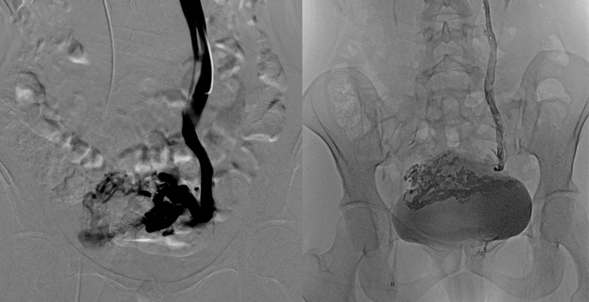
- Navigation within the veins: The catheter is guided under radiological control (usually fluoroscopy) to the dilated pelvic veins responsible for the varices.
- Phlebography: injection of iodinated contrast medium (as for a CT scan) is used to obtain precise mapping.
- Embolisation: Once the catheter is correctly positioned, embolisation material is injected into the veins to obstruct them and stop blood flow (biological glue or sclerosant).
Why undergo embolisation?
Pelvic varices embolisation is generally highly effective (in approximately 85% of cases) in reducing pain and improving patients' quality of life. Results are often visible after a few months, with a significant reduction in symptoms for the majority of patients.
Risks and side effects
Although embolisation is a relatively safe procedure, it carries certain risks, including:
- Infection: Although rare, there is a risk of infection at the catheter insertion site.
- Haematoma: Bruising may occur at the site where the catheter was inserted.
- Allergic reactions: Rarely, patients may be allergic to the materials used for embolisation.
- Symptom recurrence: In rare cases, varices may reform or symptoms may persist, requiring additional treatment.
- Post-procedural pain: Discomfort may persist for up to one month after embolisation.
Post-operative follow-up
- Patients may resume light activities after a few days.
- It is recommended to avoid strenuous physical exertion for 15 days.
- A 15-day sick leave will be prescribed.
- Imaging follow-up (MRI) is performed at 3 months, along with a consultation with the interventional radiologist to assess treatment efficacy.
How to book an appointment in Bordeaux?
Your GP or gynaecologist may refer you to the Centre Aquitain d'Imagerie in Mérignac or the Clinique Mutualiste in Pessac.
You will then be seen in consultation by an interventional radiologist who will assess with you whether intervention is necessary.
This type of procedure is covered by health insurance and your supplementary insurance.
For more information, you can visit the patient association website: https://www.info-congestionpelvienne.fr/
Ready to book an appointment?
Contact us to discuss your situation and schedule your consultation
Book an appointment